

Neck Enlargement
The neck contains numerous structures—including organs, glands, muscles, and blood vessels—making it susceptible to various types of enlargements and tumors.There are two common causes for the appearance of an enlarged mass in the neck:- Enlargement of the cervical lymph nodes, whether due to inflammation or cancerous growth.
- An enlarged nodule or cyst in the thyroid gland.
How is a neck tumor diagnosed?
Diagnosing neck masses relies on a medical examination and a comprehensive review of the patient’s medical history. We then proceed with specific tests—such as laboratory analyses and imaging—tailored to the individual patient’s needs.Ultrasound imaging provides indispensable information regarding many types of neck masses, allowing us to differentiate between various benign and malignant tumors.If further imaging is required, we may then utilize modalities such as CT scans and MRI. We must also not overlook the role of pharyngoscopy and laryngoscopy in evaluating neck masses. These procedures enable the diagnosis of internal tumors that might otherwise manifest externally only as neck swelling—such as cancerous lymph node enlargement associated with laryngeal or nasopharyngeal cancer. They also provide insight into the size of the mass and its internal impact, such as compression of internal organs, obstruction of the larynx or pharynx, or effects on the vocal cords.In some cases, a tissue sample (biopsy) is obtained from the tumor for diagnostic purposes.Carotid Body Tumor
A carotid body tumor is a rare type of neck tumor. It arises from nerve cells located at the bifurcation of the carotid artery into its two main branches.Consequently, this tumor is closely associated with the major blood vessels of the neck, and its surgical removal often presents a significant challenge.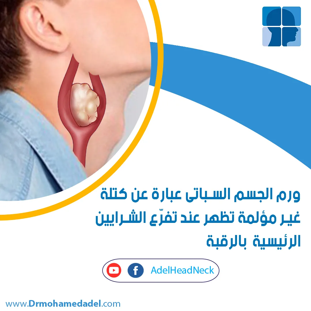
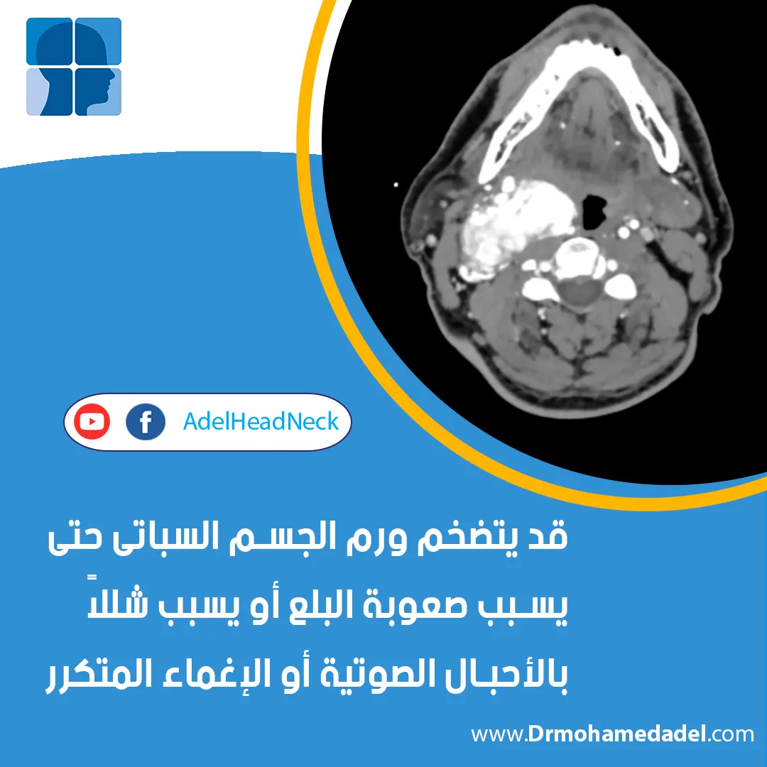
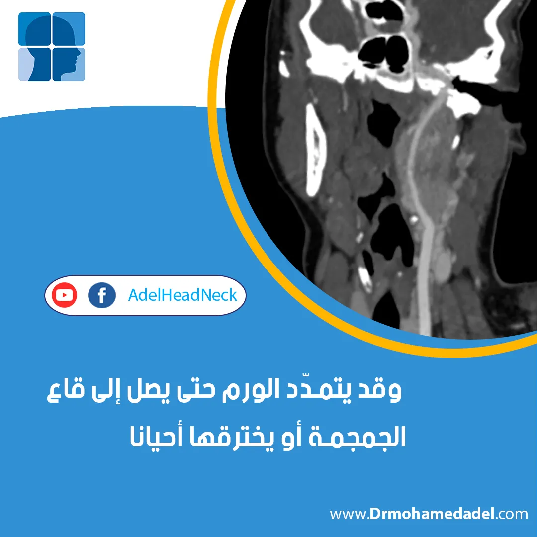
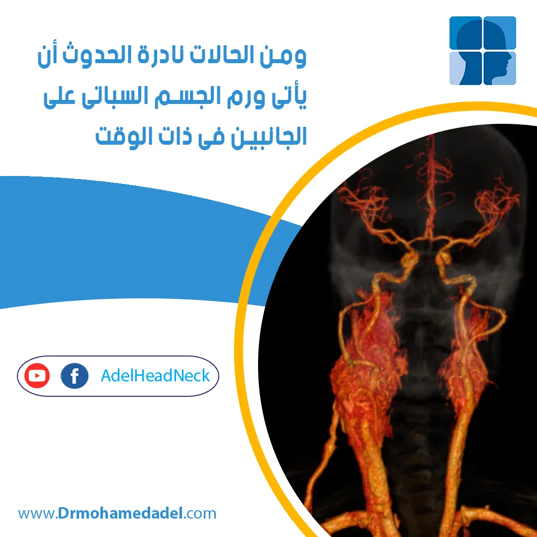
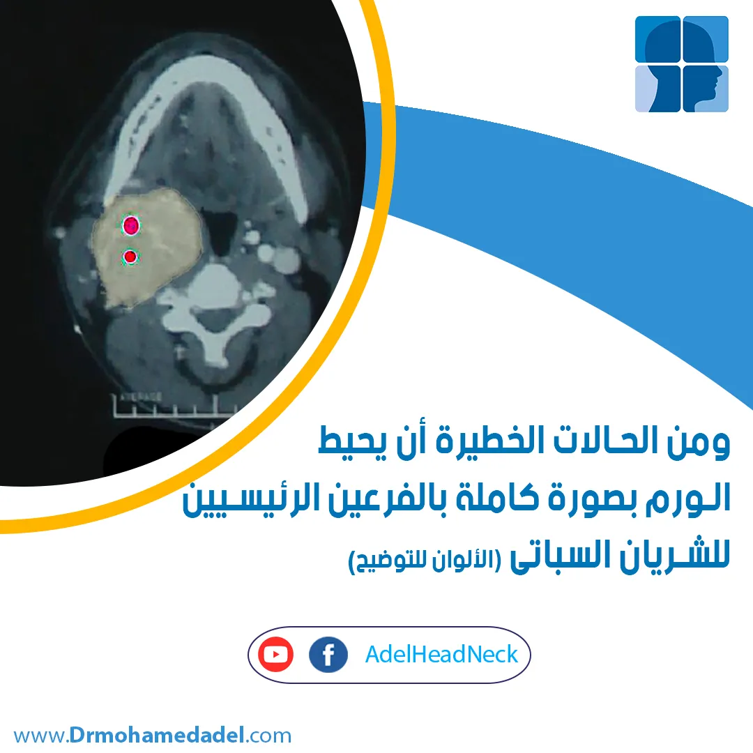

We diagnose carotid body tumors through clinical examination and imaging techniques, such as CT and MRI scans.Surgery is considered the optimal method for removing carotid body tumors; the surgeon excises the tumor while preserving the blood vessels, or performs vascular reconstruction if the tumor is densely adherent to the vessels.In some cases, we may utilize arterial catheterization and embolization prior to surgery. Arterial catheterization allows us to assess the brain’s reliance on the affected neck arteries for its blood supply. Additionally, arterial embolization helps shrink the tumor, reduce its blood supply, and minimize bleeding during the surgical procedure.
Neck Cysts
Although rare, the branchial cleft cyst is considered one of the most common types of neck cysts. It results from anatomical changes during embryonic development, leading to the formation of cysts in the neck instead of the disappearance of tissues that should have vanished as the fetus fully developed.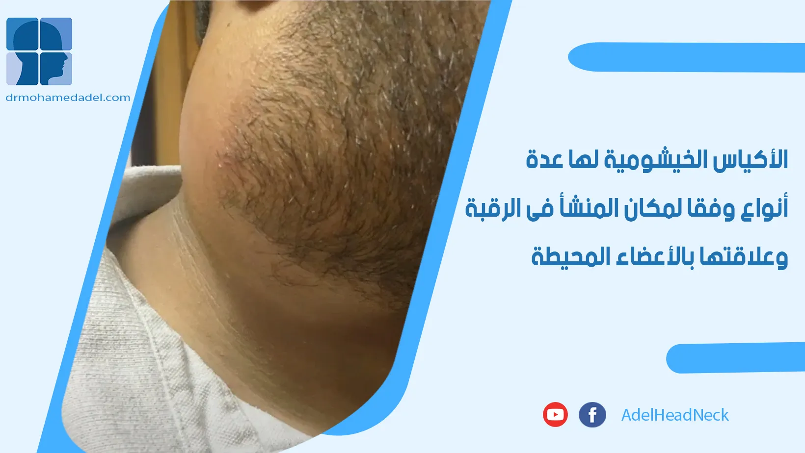

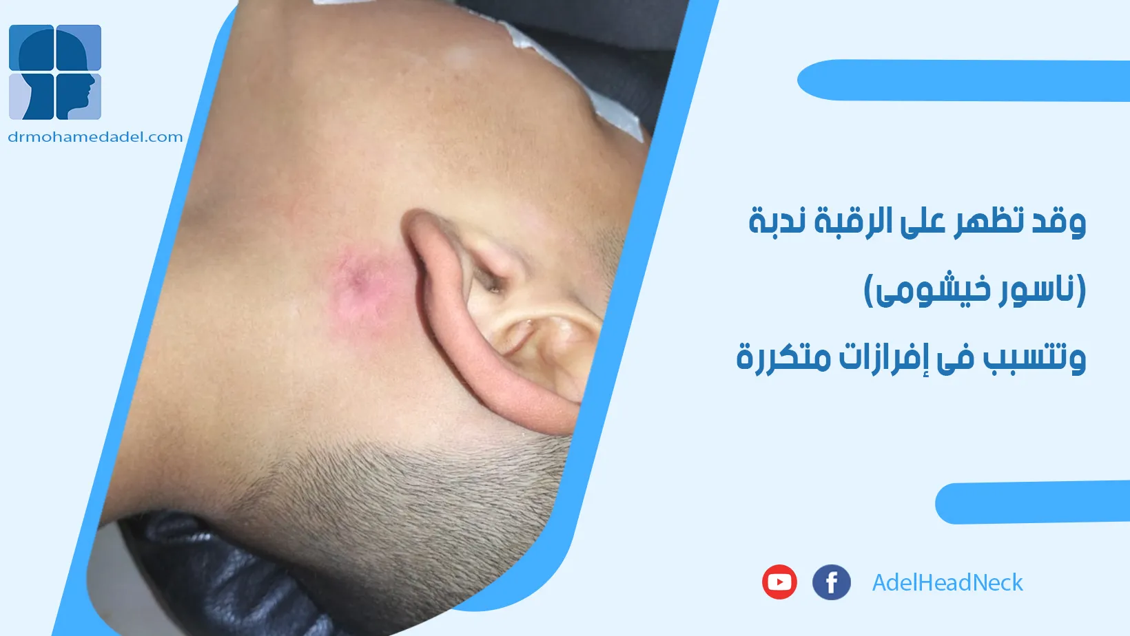
A branchial fistula presents a surgical challenge, as it may have an internal connection leading to the pharynx or the ear canal. It may also have anatomical relationships with major neck arteries or branches of the seventh cranial nerve.To learn more about branchial cysts in the neck…
The optimal approach for managing a branchial cleft cyst is surgical treatment, involving the complete excision of the cyst and any associated tract. Surgical challenges include navigating branches of the facial nerve (seventh cranial nerve) or the main carotid artery in the neck, managing critical nerves such as those controlling swallowing and tongue movement (and occasionally the laryngeal nerve), and excising any fistula or tract connecting the cyst to the throat, larynx, or tonsils.
What medical services do we offer?
Diagnostic, therapeutic, and surgical services- Diagnosis of neck masses and tumors.
- Ultrasound-guided neck biopsies.
- Surgical biopsy of cervical lymph nodes.
- Excision of neck cysts.
- Excision of neck tumors.
- Excision of branchial cleft cysts.